Exploring the Crucial Services Used by a Veterinary Cardiologist: Understanding Ultrasound and CT Scan Techniques
Vet cardiologists play a vital role in the health of pet dogs by diagnosing and treating different heart disease. They use innovative imaging methods, such as heart ultrasound and CT scans, to supply precise evaluations. Each technique has its unique benefits and applications. Understanding these techniques is important for pet dog proprietors looking for the most effective look after their companions. What factors should pet owners consider when choosing in between these diagnostic tools?

The Duty of Vet Cardiologists in Animal Health Care
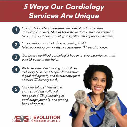
Vet cardiologists play a vital function in the healthcare of family pets, concentrating specifically on identifying and dealing with heart-related conditions. They have specialized training that enables them to analyze intricate diagnostic tests and determine various cardio issues. These specialists utilize innovative methods, such as echocardiography and electrocardiography, to examine heart function and framework accurately.Veterinary cardiologists also develop customized treatment strategies that may include medicines, lifestyle adjustments, and, sometimes, medical treatments. Their expertise includes enlightening family pet owners concerning heart wellness, stressing the value of regular examinations and early detection of potential issues. Partnership with basic veterinarians is essential, as it ensures complete take care of animals with presumed heart problems. By providing specialized solutions, vet cardiologists greatly improve the quality of life for pet dogs and provide assurance for their owners, reinforcing the value of heart wellness in overall pet dog wellness.
Common Cardiac Concerns in Pets
Typical cardiac concerns in pet dogs can considerably affect their health and lifestyle. Heart whisperings, numerous kinds of cardiomyopathy, and congenital heart issues are among one of the most widespread conditions that vets experience. CT Scans For Dogs. Comprehending these problems is essential for pet dog proprietors to ensure prompt diagnosis and appropriate treatment
Heart Murmurs in Pets
Heart murmurs can be a source of issue for pet proprietors, they are not constantly indicative of major wellness issues. A heart murmur is an abnormal noise produced by rough blood circulation within the heart. In family pets, these whisperings can be brought on by different elements, including hereditary heart issues, shutoff problems, or even stress throughout evaluations. Lots of family pets with heart murmurs lead regular lives without significant wellness impacts. To establish the underlying cause, veterinary cardiologists typically utilize diagnostic strategies such as echocardiograms and Doppler ultrasounds. Early discovery and evaluation are essential, as they might assist handle any kind of prospective heart issues properly. Family pet proprietors are encouraged to consult their vet for a complete evaluation if a heart whispering is detected.
Cardiomyopathy Kind Explained
Cardiomyopathy incorporates a group of illness affecting the heart muscle mass, bring about jeopardized heart feature in pets. The most typical types include dilated cardiomyopathy (DCM), hypertrophic cardiomyopathy (HCM), and restrictive cardiomyopathy (RCM) DCM mostly affects dogs, triggering the heart to increase the size of and damage, which lessens its ability to pump blood properly. On the other hand, HCM is a lot more prevalent in cats, identified by the enlarging of the heart wall surfaces, commonly resulting in blocked blood flow. RCM, though much less usual, happens when the heart muscle ends up being inflexible, restricting its ability to load with blood. Each type presents one-of-a-kind difficulties in diagnosis and treatment, requiring specialized veterinary cardiological evaluation to assure peak monitoring and look after influenced pet dogs.
Congenital Heart Defects
Congenital heart problems stand for a considerable category of heart concerns in animals, distinct from acquired conditions such as cardiomyopathy - CT Scans For Dogs. These flaws are architectural abnormalities existing at birth, influencing the heart's regular feature. Usual types consist of patent ductus arteriosus, ventricular septal issues, and pulmonic constriction. Symptoms may vary widely, ranging from moderate to extreme, and can include exercise intolerance, coughing, and trouble breathing. Early diagnosis via sophisticated imaging strategies like ultrasound is necessary for reliable management. Vet cardiologists play a crucial duty in recognizing these conditions and suggesting proper therapy options, which might consist of medical monitoring or surgical intervention. Acknowledging congenital heart issues enables for far better end results and enhanced high quality of life for influenced family pets
Comprehending Heart Ultrasound: Exactly How It Works
A substantial variety of veterinary methods now make use of cardiac ultrasound as an essential analysis tool for evaluating heart wellness in pets. This non-invasive method makes use of high-frequency audio waves to develop images of the heart's structure and function. Throughout the procedure, a vet professional uses a gel to the pet's breast and makes use of a transducer to send out ultrasound waves. These waves bounce off the heart and surrounding structures, producing real-time pictures on a monitor.Veterinarians can analyze various facets of heart health, including chamber size, wall motion, and valve feature. In addition, heart ultrasound enables the discovery of problems such as fluid build-up and hereditary heart flaws. This strategy is essential for diagnosing conditions that might not show up with conventional radiographs. By supplying comprehensive info about the heart's anatomy and performance, heart from this source ultrasound help in developing effective treatment prepare for pets experiencing heart disease.
The Relevance of CT Checks in Diagnosing Heart Issues
Exactly how do CT scans boost the diagnosis of heart problems in vet medicine? CT scans give in-depth cross-sectional photos of the heart and bordering structures, permitting veterinarians to picture complicated physiological connections. This imaging method is particularly beneficial in determining genetic heart flaws, heart tumors, and irregularities in capillary. By making use of advanced imaging algorithms, CT scans can examine heart chamber dimensions and function, using a complete view that may be hard to achieve with conventional methods.Additionally, CT angiography can picture blood circulation and identify locations of constriction or blockage, which is necessary for preparing prospective interventions. The speed and accuracy of CT scans likewise facilitate quick medical diagnoses, essential in emergency situations. Ultimately, the consolidation of CT scans into vet cardiology greatly improves the precision of medical diagnoses, enabling targeted treatment strategies and enhancing patient outcomes for pets experiencing heart disease.
Comparing Ultrasound and CT Scan Methods
While both ultrasound and CT scans are very useful tools in vet cardiology, they supply distinctive benefits and constraints that affect their usage in diagnosing heart conditions. Ultrasound, or echocardiography, provides real-time imaging of the heart's framework that site and function, enabling vets to evaluate heart chambers, valves, and blood flow. It is especially efficient for reviewing conditions like coronary infarction and cardiomyopathy. Ultrasound may be limited in imagining particular anatomical structures due to person dimension or obesity.In contrast, CT scans deal thorough cross-sectional pictures of the heart and bordering tissues, making them optimal for identifying structural problems, lumps, or vascular problems. CT scans supply detailed understandings, they require sedation and may entail radiation direct exposure. Inevitably, the selection in between ultrasound and CT checks depends upon the particular clinical circumstance, the patient's problem, and the details needed for an accurate medical diagnosis.
Treatment Alternatives Available With Vet Cardiology
Vet cardiology supplies a range of treatment choices tailored to address various heart disease in animals. Treatment plans often begin with way of living modifications, including diet plan changes and exercise changes, intended at enhancing overall heart wellness. Medications play an essential function, with cardiologists suggesting medicines such as diuretics, beta-blockers, and ACE inhibitors to take care of symptoms and boost heart function.In a lot more extreme instances, interventional treatments, such as balloon valvuloplasty or stent placement, may be essential to alleviate obstructions or improve blood circulation. For sure hereditary heart flaws, medical alternatives might be explored to correct structural problems. Furthermore, continuous surveillance and follow-up care are necessary parts of a complete therapy strategy, permitting prompt adjustments based on the pet's reaction to treatment. On the whole, vet cardiology concentrates on supplying efficient, personalized like enhance the health and wellness and health of pet patients with heart conditions.
Exactly how to Prepare Your Family Pet for a Heart Analysis
Preparing a pet for a cardiac examination is crucial to guarantee accurate results and a smooth process. Owners must initially set up the consultation with the vet cardiologist and discuss any type of details needs or problems. It is recommended to keep food for at the very least 12 hours before the evaluation, as this helps enhance imaging high quality throughout procedures like ultrasound or CT scans.Additionally, keeping a tranquil setting on the day of the consultation can help reduce the pet's anxiety. It is advantageous to bring along any type of appropriate medical documents, consisting of previous examinations and drugs (Cancer Veterinary Near Me). Owners ought to likewise ensure that their animal is comfy and leashed during transportation to the center. Ultimately, familiarizing themselves with the evaluation procedure can relieve worries and help in asking informed questions throughout the consultation. By following these actions, proprietors can contribute greatly to the effectiveness of the heart assessment
Regularly Asked Questions
The length of time Does a Heart Ultrasound or CT Scan Take?
The duration of a heart ultrasound generally ranges from 30 to 60 minutes, while a CT check may take around 15 to 30 mins. Variables such as the client's condition can affect these time estimates.

Exist Any Type Of Risks Connected With These Diagnostic Treatments?
Can I Stick With My Animal During the Procedure?
The vet center's plan normally dictates whether pet proprietors can continue to be during procedures. While some facilities motivate owner presence for convenience, others might need separation to assure safety and security and optimal conditions for analysis imaging.
Just how much Do These Analysis Tests Generally Price?
The expenses of diagnostic examinations, such as ultrasound and CT scans, typically differ based on area and facility. Typically, rates range from a couple of hundred to over a thousand bucks, reflecting the intricacy and technology entailed.
What Is the Recovery Refine After a Cardiac Analysis?
The recovery procedure after a cardiac examination involves checking the pet for any kind of immediate reactions, guaranteeing convenience, and limiting physical task. Veterinarians normally offer post-evaluation instructions to direct pet dog owners throughout this important recuperation period. Heart murmurs, numerous types of cardiomyopathy, and genetic heart defects are amongst the most widespread conditions that vets experience. A heart whispering is an abnormal noise generated by rough blood circulation within the heart. Cardiomyopathy encompasses a group of conditions affecting the heart muscle mass, leading to compromised cardiac feature in pet dogs. Congenital heart flaws stand for a considerable group of cardiac problems in pet dogs, distinct from gotten conditions such as cardiomyopathy. Ultrasound, or echocardiography, supplies real-time imaging of the heart's structure and function, enabling vets to assess heart chambers, valves, and blood circulation.